Advances in dairy Research
Open Access
ISSN: 2329-888X
ISSN: 2329-888X
Research Article - (2018) Volume 6, Issue 2
The aim of this study was to determine the frequency of brucellosis contagion of female calves born from seropositive and seronegative cows in brucellosis-infected herds. Brucellosis was monitored by serological analysis of 192 female calves from ten stables. Eight of these stables corresponded to family dairy herds, which had been vaccinated with S19; one was a semi-intensive herd vaccinated with RB51 and the last one was an intensive herd vaccinated with both vaccines. Monthly blood samples were taken from the female calves, from birth up to nine months of age; later, blood samples were taken at 12, 15, 18, 21, 24 months, and during delivery or abortion. Blood samples were analysed with Rose Bengal, rivanol and radial immunodiffusion tests to detect the appearance of seropositivity. A total of 192 female calves were evaluated and only 23% (45/192) were seropositive to brucellosis during the entire study. Of the 45 serologically positive female calves, 47% (21/45) were daughters of seropositive cows, while 53% (24/45) were daughters of seronegative cows. In conclusion, only 23% of the female calves born from infected herds showed seropositivity to brucellosis from birth up to 24 months of age, of which less than half were daughters of seropositive mothers and more than half were daughters of seronegative mothers. Therefore, serological diagnosis of brucellosis should be considered at an early age, which is currently not regular practice. In addition, biosecurity measures should be established, mainly in family dairy herds.
Keywords: Brucellosis; Bovine; Vaccination
Bovine brucellosis is an infective contagious disease of bacterial origin produced by Brucella abortus . It is currently of worldwide importance because it causes great economic loss to livestock industry due to a reduction in the productive and reproductive indices of infected herds. In Mexico, the disease is considered endemic, except in the State of Sonora where it has been eradicated. B. abortus transmission can occur by ingestion of food or water contaminated with the remains of abortions and/or vaginal secretions from cows infected with brucellosis, or by contact with aborted fetuses, newborn calves, placental membranes, uterine fluids or vaginal discharges of infected animals. Even in the absence of abortion, large amounts of bacteria are eliminated at delivery [1-4].
Vertical transmission occurs in 60% to 70% of animals born from infected mothers. Calves can be infected during delivery or by the intake of colostrum and unpasteurized milk from infected mothers [3,4]. Females infected in this way show no apparent signs until the reproductive age. Then they are prone to abortion during the last third of the gestation; however, in case of delivery, calves are born weak and with high probabilities of death at an early age [4-6].
Once the bacterium is established in the animal, it migrates to the local lymph nodes, where it replicates intracellularly in phagocytic cells. When this barrier is passed, the bacterium spreads along the lymphatic and blood vessels causing bacteremia, which then leads to systemic infection with lodging in the liver, spleen, bone marrow and kidneys.
The presence of erythritol is essential for B. abortus as source of carbon and energy, in order to favor survival of the microorganism in the gravid uterus of pregnant females [7,8]. The host-pathogen interaction is established in the gravid uterus [4]. Latent infection by B. abortus can be caused by introduction of infected animals into healthy herds [9].
Herds with high brucellosis prevalence are an important spreading source; once an infected cow contaminates the herd’s pen, B. abortus can remain viable for long periods if adequate temperature and humidity levels are present. The microorganism becomes even more resistant in the presence of organic matter [2-4]. In Mexico the standard against brucellosis, NOM-041, 1995, establishes serological tests for disease diagnosis, which allows determining herd status [2,10,11]. Another important serological technique to differentiate truly infected animals is the radial immunodiffusion (IDR) test; however, IDR has not yet been approved by the campaign against brucellosis.
Nevertheless, several studies show its capacity to differentiate infected organisms from vaccinated and revaccinated animals, regardless of the brucellosis strain: S19 or RB51 [12-14].
The campaign against brucellosis aims to monitor the enforcement of sanitary measures in production units. Seropositive animals must be eliminated and vaccination programs installed, and both producers and technical personnel must be trained to control the disease.
Currently, two types of vaccines are used: The B. abortus S19 strain, which has smooth morphology and allows development and persistence of post-vaccine antibodies in serum; and the B. abortus RB51 strain, a rough mutant of the 2308 strain that does not induce antibodies [3,15-17].
The aim of the study was to determine the transmission of this disease in brucellosis-infected herds to female calves born from seropositive and seronegative mothers, from birth until their first delivery or abortion.
The study was conducted with cows of ten stables, in which three different production systems are employed: eight stables were family dairy farms where herds were vaccinated with S19; one stable housed a semi-intensive herd vaccinated with RB51; and one stable kept an intensive herd and used both vaccines (Table 1). Reported brucellosis prevalence was 20% to 30%.
| Production system | Characteristics | Vaccination for brucellosis |
|---|---|---|
| Family | Eight family dairy herds, located in the municipality of Juventino Rosas, Guanajuato; with averages of 50 to 200 animals, with deficient biosecurity measures. | S19. Once in their life at 3-4 months of age. |
| Semi-intensive | A semi-intensive herd from the municipality of León, Guanajuato; with approximately 250 animals and an adequate biosecurity control. | RB51. Once in their life at 3-4 months of age. |
| Intensive | An intensive herd located in the town of El Colorado, Querétaro, with about 2,000 animals and strict biosecurity measures. | Combination of both vaccines; S19 at 3-4 months of age and one month later, revaccination with RB51. |
Table 1: Description of the three production systems that grouped the studied herds.
A total of 188 cows gave birth to female calves; four of these had female/female twin births, thus totaling 192 female calves, distributed as follows: 94 were born in family dairy herds, 28 in the semi-intensive herd and 70 in the intensive herd.
Blood samples were collected from the mother cows before calving, and examined with the Bengal Rose (RB), rivanol (RT) and radial immunodiffusion (IDR) serological tests to separate them into two categories: seropositive and seronegative mothers.
The 192 female calves born from these mothers were sampled monthly from the first to the ninth month of life. They were then sampled at 12, 15, 18, 21, 24 months of age, until either calving and/or abortion occurred.
Serological tests were performed to determine the time at which the serological reaction was first positive. Seropositivity to brucellosis was considered when female calves showed a positive reaction to the BR and RT tests, starting at birth and before vaccination.
After vaccination, production system family and intensive were tested with IDR in addition to the BR and RT tests in order to differentiate vaccine antibodies from infection antibodies.
Thus, female calves positive to brucellosis showed a reaction to the three tests. In semi intensive positive female calves presented serum agglutination to the BR and RT tests.
To approximate the age at which calves were serologically positive to brucellosis in the considered categories (i.e. daughters of seropositive and seronegative cows), only the month was considered in which the positive reaction appeared in the respective tests.
Thus, only new cases were considered at each sampling. Frequency data analysis was performed to determine the time at which antibodies of B. abortus appeared in the two categories.
Of the 188 mother cows evaluated by BR and RT tests (mothers of female calves of the three production systems), 23% (44/188) were seropositive, and 77% (144/188) were seronegative for brucellosis.
A total of 192 female calves were tested for seropositivity along a 24- month period; these calves were daughters of either seropositive or seronegative mothers from the three production systems. Results showed that only 23% (45/192) of evaluated female calves were seropositive to brucellosis.
Of the 45 serologically positive female calves, 47% (21/45) were daughters of seropositive cows and 53% (24/45) were daughters of seronegative cows (Graph 1).
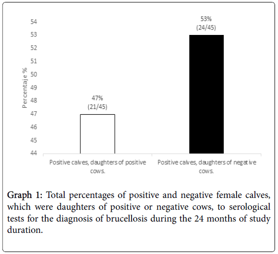
Graph 1: Total percentages of positive and negative female calves, which were daughters of positive or negative cows, to serological tests for the diagnosis of brucellosis during the 24 months of study duration.
In female calves, daughters of seropositive cows, seropositivity appeared in 81% (17/21) during the first 30 days of life (Graph 2).
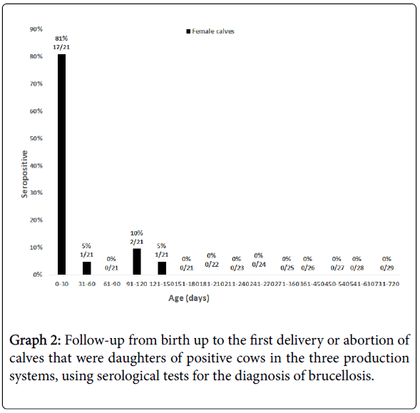
Graph 2: Follow-up from birth up to the first delivery or abortion of calves that were daughters of positive cows in the three production systems, using serological tests for the diagnosis of brucellosis.
During the 24 months of follow-up of seropositive calves (daughters of seronegative cows), we observed variability in most of the samples. The highest percentage of seropositivity was obtained at 120 days of age (i.e. 33%; 8/24). However, seroconversion was observed in this category at the time of delivery or abortion (i.e. 13%; 3/24) (Graph 3).
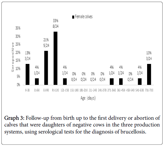
Graph 3: Follow-up from birth up to the first delivery or abortion of calves that were daughters of negative cows in the three production systems, using serological tests for the diagnosis of brucellosis.
Compared by production system, during the 24 months of the follow-up, the family herd system lodged the highest number of seropositive calves, daughters of both seropositive and seronegative cows, corresponding to 90% (19/21) and 63% (15/24), respectively (Graph 4).
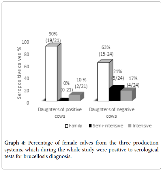
Graph 4: Percentage of female calves from the three production systems, which during the whole study were positive to serological tests for brucellosis diagnosis.
In Mexico, bovine brucellosis has a direct and important impact on livestock production as the disease is endemic almost throughout the country. In female livestock, brucellosis is characterized by a high incidence of abortions.
The Official Mexican Standard to control brucellosis focuses on serological diagnosis of adult animals, vaccination of female calves and elimination of seropositive animals. However, these control measures are not followed strictly, and, consequently, the disease has spread and remains endemic in the country.
Female calves born in herds infected with brucellosis have a risk of infection at some point of their lives, depending on the management and biosecurity measures established by the production system, indicate that continuous serological monitoring should be applied in order to identify infected cows and these should be promptly separated from the herd.
These measures are necessary to avoid infecting healthy animals at the time of calving or abortion; in addition, seropositive animals should be gradually removed from the herd once their milk production decreases or if they show any health problem, as calves can be infected by sucking on colostrum or milk, and by passing through the vaginal canal of infected mothers [4].
In a report by Carrizosa, 9.1% seropositivity to brucellosis was reported in calf daughters of positive cows during the first week and three months of age and 18.2% (4/22) of seropositive calf daughters of negative cows during the same period.
These results contrast with those from the present study, however, where the percentage of female calves that proved positive to brucellosis during the first month of life was 81% (17/21), when born from seropositive cows, and, 13% (3/24) when born from seronegative cows.
Of the total sample examined in the present study, an overall 23% of female calves were seropositive for brucellosis. Taking this as the total, 47% seropositive calves were daughters of seropositive cows and 53% were daughters of seronegative cows. The latter percentage was, in fact, quite surprising, because a lower infection risk would be expected from the daughters of seronegative cows.
This result should be stressed because it is related to the management of calves during their developmental stages. The high infection rate could be due to administration to newborn calves of colostrum or milk from seropositive cows or to the coexistence of infected and non-infected cows in the same pens [4].
Regarding the time of infection, daughters of seropositive cows showed the highest percentage of seropositivity during the first month of life (81%), while in the daughters of seronegative cows, results were variable along the 24 months of the study: seropositive calves were found at birth (13%), and in the third (21%) and fourth months of age (33%).
These results may be due to poor biosecurity measures applied at the stable where the colostrum or milk of positive and negative cows could be mixed; or the lack of a cleaning and disinfection program at the time of delivery or abortion. In addition, since brucellosis is caused by facultative intracellular bacteria [18], it is also possible that, at the time of sampling of cows with seronegative results, antibodies were not detected because these occur intermittently.
This suggests that results of infected cows may be false negative and, if so, bacteria would be transmitted by their colostrum or milk [4].
With respect to the productive management system, the highest percentage of contagion occurred in family dairy herds (76% of seropositive female calves) due to the deficiencies in health management, which allow the spread of the disease within the herd and contagion to the calves [19]. This study describes various risk factors that are associated with the transmission of bovine brucellosis in dairy herds in the state of Baja California, such as: the purchase of replacements, the presence of dogs, and the mix of healthy cows with seropositive animals during milking, inadequate handling of the calving or abortion wastes, and not eliminating animals positive to brucellosis.
On the other hand, the semi-intensive herd (11% of seropositive female calves) had management control but it was incomplete, since seropositive animals were identified and separated, but at the time of delivery (or abortion) all cows were confined to a single pen, allowing the infection to spread among the animals [20].
In the intensive system, in spite of having strict biosecurity control measures of seropositive adult animals, 13% of seropositivity occurred. This management does not apply to female calves; thus, when they reach adulthood there is still a risk factor for healthy animals at the time of delivery or abortion.
In a study conducted by Herrera, he reported that the milk increase in the herd was proportional to the reduction of new cases of brucellosis; so, he showed that it is important to consider the elimination of seropositive animals, and elimination should be done at an early age, once they are diagnosed as positive to bovine brucellosis.
It the present study, only 23% of female calves born from infected herds were seropositive to brucellosis, monitored from birth to 24 months of age [21]. Of these calves, 47% corresponded to daughters of positive cows and 53% to daughters of negative cows. The most representative seropositivity period in both categories was before the first month of life.
In conclusion, it is important to consider the serological diagnosis at an early age; especially in family dairy herds with few biosecurity measures, where the risk of contracting the disease is high.
This work was partially supported by Guanajuato Produce A.C Foundation: Project FGP601-13 “Technology transference in sanitary aspects of a family dairy in the state of Guanajuato”.