Journal of Clinical & Experimental Dermatology Research
Open Access
ISSN: 2155-9554
ISSN: 2155-9554
Case Report - (2020)Volume 11, Issue 5
Fixed drug eruption (FDE) is an adverse drug reaction characterised by recurrent plaques within the same site after drug ingestion. The typical pigmented evolution and notion of drug intake have considerable diagnostic value. Eyelid is a rare localization of FDE that may be challenging for diagnosis, especially when allergological testing is not possible. The risk of further adverse reaction and ocular involvement makes it necessary for physicians to be familiar with this localization. We hereby report a case of a solitary FDE of the left upper eyelid.
Fixed drug eruption; Eyelid; Piroxicam
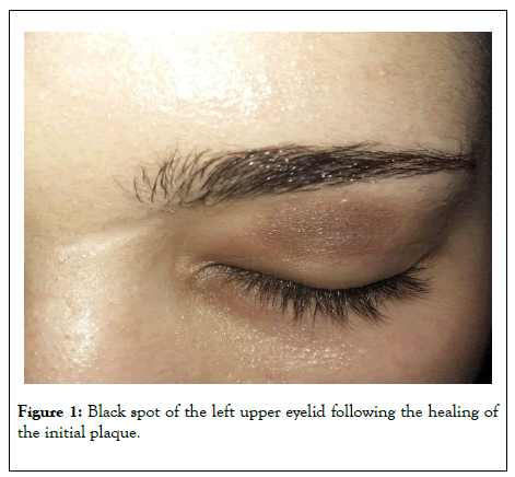
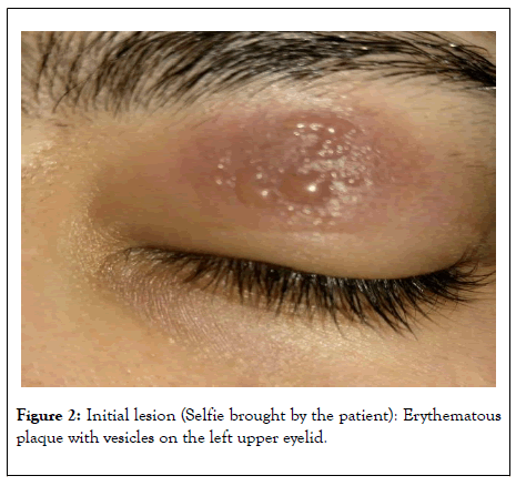
A 23 years old woman with a history of chronic dysmenorrhea requiring frequent use of NSAIDs, presented with a pigmented upper left eyelid spot that appeared two weeks earlier. The patient reported taking Piroxicam in self-medication 3 days before the installation of an erythematous and painful plaque of the left upper eyelid evolving towards a pigmented spot (Figure 1). A selfie picture taken by the patient at this moment revealed a rounded well-defined erythematous plaque surmounted by vesicles (Figure 2). The diagnosis of bullous fixed drug eruption with Piroxicam was made on the basis of clinical and chronological arguments. Avoidance of Proxicam and other related NSAIDs was the main treatment since no specific therapy could be prescribed owing to the late stage of the pigmented evolution. No further episodes were observed.

Figure 1: Black spot of the left upper eyelid following the healing of the initial plaque.

Figure 2: Initial lesion (Selfie brought by the patient): Erythematous plaque with vesicles on the left upper eyelid.
Fixed drug eruption (FDE) is characterized by the occurrence, upon ingestion of a drug, of recurrent erythematous plaques within the same skin or mucosal site, and a suggestive pigmented evolution. NSAIDs are frequently incriminated but only few cases due to piroxicam have been reported so far in literature [1]. Classical clinical presentation is made of single or multiple cutaneous and/or mucosal lesions of variable topography but mainly located on the face and trunk. The association of a specific localization to a particular drug has not been yet established and remains controversial, but Lips seem to be an important site for oxicam-induced FDE [2]. Single bullous localization to the eyelid is not evocative and has never been reported with piroxicam. Kimmatkaar et al [3] reported a challenging case of solitary paracetamol-induced FDE of the eyelid presenting as skin necrosis and misleading the diagnosis toward a necrotizing fasciitis. For our patient alternative diagnoses considered were eczema, insect bite, herpetic infection and lichen pigmentosa. The notion of drug intake few days before the occurrence of the lesion was very important to query in such non suggestive case. Anatomical characteristics of the eyelid, especially the thinness of the integument, could possibly explain the atypical presentations within this area, as necrotic aspects [3] or vesiculation observed in our patient. Other ocular symptoms may include conjunctival edema and injection [4].
In our case Patch tests, oral provocation and skin biopsy were considered unnecessary owing the low risk to benefit ratio, and the typical clinical history. Discontinuation of the causative drug is the regular treatment of FDE, but patients with piroxicaminduced FDE should also avoid other oxicams because of possible cross sensitivity [5]. Topical steroids may have a benefit on the pigmentation when used at the early stage of the disease [6].
Isolated palpebral involvement is a rare localization of FDE which has been poorly described. The risk of bullous evolution in case of reintroduction of the causative drug and the aesthetic sequelae, emphasizes the necessity for dermatologists and ophthalmologists to be familiar with this localization and to evoke the diagnosis in front of any pigmented lesion of the eyelid.
Citation: Khallaayoune M, Sialiti S, Elgaitibi F, Ismaili N (2020) Single Localization of Bullous Fixed Drug Eruption on the Eyelid: An Unusual Presentation for a Classic Diagnosis. J Clin Exp Dermatol Res. 11:533. DOI: 10.35248/2155-9554.20.11.533
Received: 12-Aug-2020 Accepted: 26-Aug-2020 Published: 31-Aug-2020 , DOI: 10.35248/2155-9554.20.11.533
Copyright: © 2020 Khallaayoune M, et al. This is an open-access article distributed under the terms of the Creative Commons Attribution License, which permits unrestricted use, distribution, and reproduction in any medium, provided the original author and source are credited.