Mycobacterial Diseases
Open Access
ISSN: 2161-1068
ISSN: 2161-1068
Case Report - (2023)Volume 13, Issue 2
The sporotrichoid distribution of lesions has been described in sporotrichosis, lupus vulgaris, atypical mycobacteria, chromoblastomycosis, nocardia, cat-scratch disease. Apart from these infectious diseases some other conditions like melanoma, squamous cell carcinoma, B cell lymphoma and T cell lymphoma, epithelioid sarcoma, and peripheral nerve sheath tumor had also showed sporotrichoid spread. A 45 year old otherwise healthy male presented with mild painful skin colored to erythematous plaque, initially started over the left dorsa of the ankle, later new plaque appeared to involve the left leg and thigh linearly with an increase in thickness and size of plaque over a period of 4 months. On examination typical sporotrichoid distribution of three well defined, minimally tender edematous, indurated, skin colored to an erythematous plaque with central clearing in large plaque with overlying fine white collarette, non-adherent scale with no sinus, scarring, verrucosity, pustules over left ankle. Based on history and examination differentials of lymphocutaneous sporotrichosis, sporotrichoid lupus vulgaris, wells syndrome, Borderline Tuberculoid (BT) Hansen with resolving type 1 reaction were kept. Biopsy from the erythematous plaque reveals oblong well-defined granuloma composed of epithelioid cells, dense eosinophils. Evaluation for hypereosinophilia yielded no results (stool for ova cyst, chest x-ray, cardiac echo, and serum Immunoglobulin E (IgE) were normal). Though acid-fast stain for lepra bacilli was negative, a final diagnosis of borderline tuberculoid Hansen with resolving type 1 reaction with hypereosinophilic syndrome was made. Hansen’s disease presents with a various morphological spectrum ranging from hypopigmented macule, erythematous plaque, nodule, diffuse infiltration, ulceration, shiny dome-shaped papules and necrotic plaques. Sporotrichoid distribution has not yet been described. Till date, only single case report noted the association of hansen and hypereosinophilic syndrome, this might be any association or mere coincidence.
Hansen; Hypereosinophilic syndrome; Sporotrichoid spread; Lymphatic spread
Hansen’s disease is a chronic granulomatous disease caused by acid fast Bacillus i.e, Mycobacterium leprae, an acid fast Bacillus that lives in the cytoplasm of macrophages and Schwann cells. It usually affects the peripheral nervous system, upper respiratory system, testes, skin, and ocular involvement [1]. Hansen’s disease presents as a various morphological spectrum ranging from hypopigmented macule, erythematous plaque, nodule, diffuse infiltration, ulceration, shiny dome-shaped papules and necrotic plaques [2]. Sporotrichoid distribution has not yet been described in this study.
A 45 year old otherwise healthy male presented with mild painful skin colored to erythematous plaque, initially started over the left dorsa of the ankle, later new plaque appeared to involve the left leg and thigh linearly with an increase in thickness, size of plaque over a period of 4 months. Initially started as 2 × 2 cm plaque, it slowly progressed to the current size of 3 × 3 cm (minimum) and 6 × 4 cm (maximum). He was on continuous analgesics for arthralgia and tenderness over plaques. The patient has no history of any of the following conditions: Urticaria, angioedema, weight loss, fever, or prior trauma.
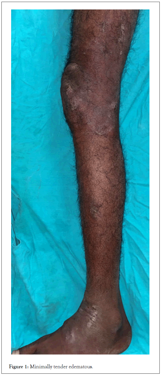
On examination typical sporotrichoid distribution of three welldefined (Figure 1) minimally tender edematous, indurated, skin colored to an erythematous plaque with central clearing in large plaque with overlying fine white collarette, non-adherent scale with no sinus, scarring, verrucosity, pustules over left ankle (Figure 2), left lateral thigh (Figure 3). No local rise of temperature, fever, weight loss, or trauma. No feeder nerve, no sensory or motor weakness, no lymphadenopathy and Systemic examination were completely normal.

Figure 1: Minimally tender edematous.
Figure 2: Pustules over left ankle.
Figure 3: Left lateral thigh.
Based on history and examination differentials of lymphocutaneous sporotrichosis, sporotrichoid lupus vulgaris, well’s syndrome, Borderline Tuberculoid (BT) Hansen with resolving type 1 reaction were kept. Biopsy from the erythematous plaque reveals oblong welldefined granuloma composed of epithelioid cells, dense eosinophils (Figure 4), and foamy histiocytic infiltrate in perivascular, peri adnexal distribution (Figure 5). Fite stain and Periodic Acid Schiff stain were negative. Tissue potassium hydroxide mount and fungal culture, tissue Cartridge-Based Nucleic Acid Amplification Test (CBNAAT) and mycobacterial culture were also showed negative results. Slit skin smear from plaque and ear lobule were also showed negative results. Dense eosinophilic infiltrate in the tissue specimen was correlated with peripheral blood eosinophilia (1600/μl). Evaluation for hypereosinophilia yielded no possible results (stool for ova cyst, chest X-ray, cardiac echo, and serum Immunoglobulin E (IgE) levels were normal). Though acid-fast stain for lepra bacilli was negative, a final diagnosis of Borderline Tuberculoid (BT) Hansen with resolving type 1 reaction with hypereosinophilic syndrome was made based on characteristic distribution of foamy histiocytic granuloma and idiopathic hypereosinophilia. The patient was started on Multi Bacillary Multi Drug Therapy (MBMDT) and oral steroids. After 4 weeks of treatment, complete resolution of plaque was noted (Figures 6 and 7).
Figure 4: Epidermal acanthosis, underlying oblong well-defined granuloma composed of epithelioid cells, foamy histiocytic infiltrate in perivascular, peri adnexal distribution.
Figure 5: Epidermal acanthosis, underlying granuloma composed of epithelioid cells, foamy histiocytic infiltrate.
Figure 6: The patient was started on Multi Bacillary Multi Drug Therapy (MBMDT) and oral steroids.
Figure 7: After 4 weeks of treatment, complete resolution of plaque.
The sporotrichoid distribution of lesions has been described in sporotrichosis, lupus vulgaris, atypical mycobacteria, chromoblastomycosis, leishmaniasis, histoplasmosis, cryptococcosis, cowpox infection, nocardia, cat-scratch disease [3]. A part from these infectious diseases some other conditions like melanoma [4], Bowen’s disease, squamous cell carcinoma [5], B cell lymphoma and T cell lymphoma [6], epithelioid sarcoma, and peripheral nerve sheath tumor shows sporotrichoid spread [7]. Hansen’s disease presenting in this fashion has not yet been described in this study. As we know, lymphadenopathy is seen in Hansen’s disease as a direct invasion or as a reactive process. Similar to lymph node involvement may be a spill of lepra bacilli in the lymphatics results in sporotrichoid distribution of Hansen’s disease as in our case.
Hypereosinophilic syndrome is described when peripheral blood eosinophilia (more 1500/µL), absence of an underlying cause, organ damage as a result of presence of toxic eosinophilic contents. Major tissue targets are skin, central nervous system, heart and lung. Cutaneous manifestations described are urticaria, angioedema, erythematous pruritic papules and nodules. It presents as wide spectrum ranging from mild disease requiring no treatment (as in our case) to fatal outcome due to cardiac involvement. Treatment includes high dose glucocorticosteroids, if features of churg strauss is suspected then cyclophosphamide is added. Treatment with methotrexate, cyclosporine, interferon alfa, hydroxyl urea, and imatinib are used in systemic involvement [8]. Till date only a single case report explaining the Hansen with Hypereosinophilic syndrome was described. This is just a mere coincidence or any association need to be further explored [9]. The distribution, history of subsequent appearance of plaque in sporotrichoid fashion, and the presence of eosinophils in granuloma were unusual in Hansen’s disease. This case emphasizes that even Hansen’s disease can present as sporotrichoid manner. Sporotrichoid spread warrants good clinicopathological correlation in diagnosing the condition and initiation of appropriate treatment. Further reports and observation are needed to know whether the hypereosinophilia as in our is just a coincidence or work-up for underlying parasitic manifestations in the gut or elsewhere or primary eosinophilia.
This study emphasizes that even Hansen’s disease can present as sporotrichoid manner. Sporotrichoid spread warrants good clinicopathological correlation in diagnosing the condition and initiation of appropriate treatment. Hansen’s disease presents as a various morphological spectrum ranging from hypopigmented macule, erythematous plaque, nodule, diffuse infiltration, ulceration, shiny dome-shaped papules, necrotic plaques.
[Crossref] [Google Scholar] [PubMed]
[Google Scholar] [PubMed]
[Crossref] [Google Scholar] [PubMed]
[Google Scholar] [PubMed]
[Crossref] [Google Scholar] [PubMed]
[Crossref] [Google Scholar] [PubMed]
[Crossref] [Google Scholar] [PubMed]
[Google Scholar] [PubMed]
Citation: Shreya K, Sahni MK, Panwar H, Rokde R, Asati DP (2023) Sporotrichoid Hansen’s disease with Hypereosinophilic Syndrome: A Clinicopathological Challenge. Mycobact Dis. 13:322.
Received: 01-Feb-2023, Manuscript No. MDTL-23-22023; Editor assigned: 03-Feb-2023, Pre QC No. MDTL-23-22023 (PQ); Reviewed: 17-Feb-2023, QC No. MDTL-23-22023; Revised: 24-Feb-2023, Manuscript No. MDTL-23-22023 (R); Published: 03-Mar-2023 , DOI: 10.35248/2161-1068.23.13.322
Copyright: © 2023 Shreya K, et al. This is an open-access article distributed under the terms of the Creative Commons Attribution License, which permits unrestricted use, distribution, and reproduction in any medium, provided the original author and source are credited.