Drug Designing: Open Access
Open Access
ISSN: 2169-0138
ISSN: 2169-0138
Research Article - (2016) Volume 5, Issue 1
The causative agent of the guinea worm disease – Dracunculus medinensis, which is the only species that infects human. The intermediate host Cyclops (the small water fleas) ingest the larvae of parasite (D. medinensis) which is further ingested by the human from the stagnant contaminated unfiltered water from the source. Soon after ingestion the Cyclops is disintegrated by stomach digestive juices and causes the release of the larvae. These larvae travel and penetrate the digestive wall into the body cavity and get entry in abdominal cavity and retroperitoneal space. These larvae mature in adults and soon after the copulation the ovoviviparous female mature and grows in size whereas the male dies. After incubation period of a year or year and half the mature female worm come towards the skin and start formation of a small round bulge on the skin by secreting an irritating chemical. This blister is the first sign of identification that a person has got infected by guinea worm. NADH dehydrogenase subunit 5 (mitochondrion ) protein of the D. medinensis is a 527 a protein which is used for the identification of the antigenicity through B- cell epitopes prediction methods. The result obtained shows that the region of maximal hydrophilicity is likely to be antigenic site having the hydrophobic characteristics and contain the segments of low complexity and high-predicted flexibility. This predicted antigenic protein from D. medinensis could be the new paradigm of synthetic vaccine development and target validation.
<Keywords: Antigen; Dracunculusmedinensis; Epitope; Protein; Vaccine; NADH dehydrogenase subunit 5 (mitochondrion)
In this study NADH dehydrogenase subunit 5 (mitochondrion) protein has been used to investigate its role in antigenicity. NADH dehydrogenase subunit 5 (mitochondrion) protein is an active protein in mitochondria and it is a part of a large enzyme complex known as complex I. Complex I play an important role in oxidative phosphorylation. The NADH dehydrogenase complex (or mitochondrial respiratory complex I) catalyzes the oxidation of NADH by ubiquinone. This reaction is linked to proton transfer across mitochondrial membranes [1]. Mutation in the mitochondrial NADH dehydrogenase subunit 5(MT-ND5) causes mitochondrial encephalomyopathy, lactic acidosis, and strokelike episodes (MELAS), Leigh’s syndrome and Leber’s hereditary optic neuropathy. It has been found that mitochondrial ND5 12338T>C variant is generally associated with maternally inherited hypertrophic cardiomyopathy in a Chinese pedigree and this investigation suggest that thetheoretical basis for genetic counseling is required for maternally inherited hypertrophic cardiomyopathy [2]. The mutation of the mitochondrial ND5 T12338C is found to be associated with Leber’s hereditary optic neuropathy which has been investigated in the two chinese families [3]. Investigation reveals that the mitochondrial ND5 as the causative gene of Leight syndrome [4]. Guinea worm is the largest tissue parasite which is the only species that infect humans. It has got very unusual life cycle with incubation period of the approximately more than a year with six developmental stages. This one of the most neglected tropic parasite which bears clinical importance and needs to be eradicated after small pox [5]. Mature and adult female after the copulation produces millions of eggs in its uterus, and is predominantly localized in the lower extremities (80-90%). After an incubation period the female worm release the larvae which induces a painful blister (1 to 6cm diameter ) on the skin of lower limbs; the person developa slight fever , localskinredness , swelling and severe pruritus around the blister . Other symptoms include diarrhea, nausea, vomiting and dizziness. The blister burst within 1 to 3 days and female worms one or more slowly comes out from the wounds which causes an excoriating burning sensation and pain [6]. Immersing or pouring water over the blister provide pain reliever. But this the moment that adult female is exposed to the external environment [7]. Duringemergence of the limbs in open water sources it recognizes the temperature difference and releases the milky white liquid in the water which contains millions of immature larvae, when larvae released in water are ingested by copepods where they mount twice and become infective larvae within two weeks [8]. The D. medinensis antigen peptides can be most desirable segment for the subunit vaccine development because with the single epitope, the immune response can be generated in large population. This approach is usually based on the phenomenon of cross-protection, whereby infected with the mild strain and is protected against a more severe strain of the same. The phenotype of the resistant transgenic hosts includes fewer centers of initial infection, a delay in symptom development and low accumulation. B-cell epitopes are the sites of molecules that are recognized by antibodies of the immune system. Knowledge of B-cell epitopes may be used in the design of vaccines and diagnostics tests. It is therefore of interest to develop improved methods for predicting B-cell epitopes [9]. Antigen protein prediction from D. medinensis is necessary for few paradigms of synthetic vaccine development and target validation [10,11].
Database searching
The protein sequence of NADH dehydrogenase subunit 5 (mitochondrion) from Dracunculus medinensis was retrieved from www.ncbi.nlm.nih.gov, UniProt databases are initially the most important step [12,13].
Prediction of antigenicity
Prediction of antigenicity program predicts those segments from NADH dehydrogenase subunit 5 (mitochondrion) protein that are likely to be antigenic by eliciting an antibody response. In this research work antigenic epitopes of Dracunculus medinensis- NADH dehydrogenase subunit 5 (mitochondrion) are determined by using the Welling, Parker, Bepipred , Kolaskar and Tongaonkar antigenicity methods [14-22].
Solvent accessible regions
We also analyzed the solvent accessible regions of proteins having highest probability that a given protein region lies on the surface of a protein Surface Accessibility, backbone or chain flexibility by Emini et al. [23] and Karplus and Schulz [24]. By using different scale we predict the hydrophobic and hydrophilic characteristics of amino acids that are rich in charged and polar residues residues i.e., Kyte and Doolittle [25], Abraham and Leo [26], Bull and Breese [27], Miyazawa et al. [28], Roseman [29], Wilson et al. [30], Cowan [31].
Prediction of antigenic peptides
In this study, we have used bioinformatics tools approach to found the antigenic determinants. The Welling antigenicity plot gives value as the log of the quotient between percentage in a sample of known antigenic regions and percentage in average proteins and prediction result data found high in Position: 257, Score: 0.742 (max) i.e., 254-KLVALST-260 (Figure 1). We also study Hydrophobicity plot of HPLC / Parker Hydrophilicity prediction result data found in position : 308(residue: Q) i.e., 305-GGQQDSR-311 and in position 309(residue: D) i.e 306-GQQDSRG-312 with maximum score:6.3. (Figure 2 and Table 1), BepiPred predicts the location of linear B-cell epitopes result found in position: 309 (residue: D) with maximum score:1.369 i.e., 303-GYIIH LC-312 (Figure 3 and Table 2), Kolaskar and Tongaonkar antigenicity methods (Figure 4 and Table 3) predicted peptides result found in position 449 (residue: C) with high score 1.307 i.e., 446-LVLCVVF-452. The highest score for the residue indicates the probability to be a part of the epitope (Residue colored in yellow). There is the probability that the predicted antigenic fragments can bind to MHC molecule is the first bottlenecks in drug design.
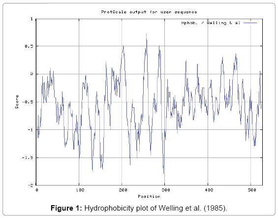
Figure 1: Hydrophobicity plot of Welling et al. (1985).
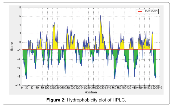
Figure 2: Hydrophobicity plot of HPLC.
| Prediction Method | Predicted residue scores | |||||
|---|---|---|---|---|---|---|
| Kolaskarand Tongaonkar Antigenicity Prediction | Position | Residue | Start | End | Peptide | Score |
| 449 | C | 446 | 452 | LVLCVVF | I.307 | |
| 407 | V | 404 | 410 | VSLVLVV | 1.292 | |
| 145 | L | 142 | 148 | CVFLVFC | 1.289 | |
| 144 | F | 141 | 147 | VCVFLVF | 1.285 | |
| 450 | V | 447 | 453 | VLCVVFF | 1.285 | |
| 448 | L | 445 | 451 | GLVLCVV | 1.276 | |
| 56 | V | 53 | 59 | CLLVMVV | 1.27 | |
Table 1: Kolaskar and Tongaonkar antigenicity prediction table.
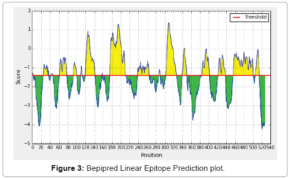
Figure 3: Bepipred Linear Epitope Prediction plot.
| Prediction Method | Predicted residue scores | |||||
|---|---|---|---|---|---|---|
| Parker Hydrophilicity Prediction | Position | Residue | Start | End | Peptide | Score |
| 308 | Q | 305 | 311 | GGQQDSR | 6.3 | |
| 309 | D | 306 | 312 | GQQDSRG | 6.3 | |
| 307 | Q | 304 | 310 | CGGQQDS | 5.9 | |
| 310 | S | 307 | 313 | QQDSRGY | 5.214 | |
| 344 | V | 341 | 347 | GGSVSKE | 4.886 | |
Table 2: Parker hydrophilicity prediction table.
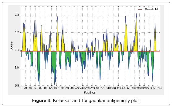
Figure 4: Kolaskar and Tongaonkar antigenicity plot.
| Prediction Method | Predicted residue scores | |||
|---|---|---|---|---|
| Bepipred Linear Epitope Prediction Prediction | Position | Residue | Score | End |
| 309 | D | 1.369 | - | |
| 308 | Q | 1.349 | - | |
| 193 | S | 1.295 | - | |
| 195 | P | 1.276 | - | |
| 194 | A | 1.228 | - | |
Table 3: Bepipred linear epitope prediction table.
Solvent accessible regions
We also predicted the solvent accessible regions in proteins; different measurement was performed for the prediction of antigenic activity, surface region of peptides. Emini et al. [23] (Figure 5) predicts the highest probability i.e. found in position: 308 (residue: Q) i.e., 306-GQQDSR-311 and in position 309 (residue: D) i.e., 307-QQDSRG-312 with maximum score:7.194, that a given protein region lies on the surface of a protein and are used to identify antigenic determinants on the surface of proteins. Karplus and Schulz (Figure 6) is found i.e.position:308 (residue: Q) i.e., 305-GGQQDSR-311 with highest score: 1.122 and in position: 307 (residue: Q) i.e., 304-CGGQQDS-310 with high score: 1.111. Predict backbone or chain flexibility on the basis of the known temperature B factors of the a-carbons. The hydrophobicity and hydrophilic characteristics of amino acids is determined by using different scales that are rich in charged and polar residues i.e. Kyte and Doolittle result high in Position: Position:14, Score: 3.667 (max)(11- ILLCFLL-17) (Figure 7), Abraham and Leo result high in Position: Position: 364, Score: 2.423 (max)(361-FLVFLFF-367) (Figure 8), Bull and Breese result high in Position: Position: 308, Score: 0.717 (max)(305-GGQQDSR-311) (Figure 9), Miyazawa result high at Position: Position: 364, Score: 8.688 (max) (361-FLVFLFF-367) (Figure 10), Roseman result high in Position: 364, Score: 2.012 (max) (361- FLVFLFF-367) (Figure 11), Wilson et al. [30] in Position: 364, Score: 7.022 (max) (361-FLVFLFF-367) (Figure 12), Cowan in Position: 364 Score: 1.716 (max) (361-FLVFLFF -367) (Figure 13). The predicted antigenic protein segments of NADH dehydrogenase subunit 5 can take active part in the host immune reactions. In future study the predicted antigenic protein NADH dehydrogenase subunit 5 fragments can be used in the investigation of MHC molecules binding and it can be the first bottlenecks in vaccine design.
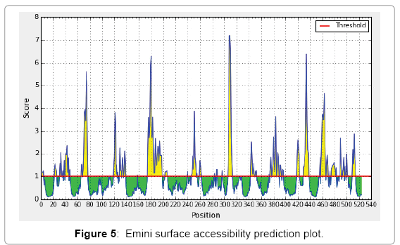
Figure 5: Emini surface accessibility prediction plot.
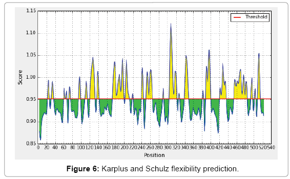
Figure 6: Karplus and Schulz flexibility prediction.
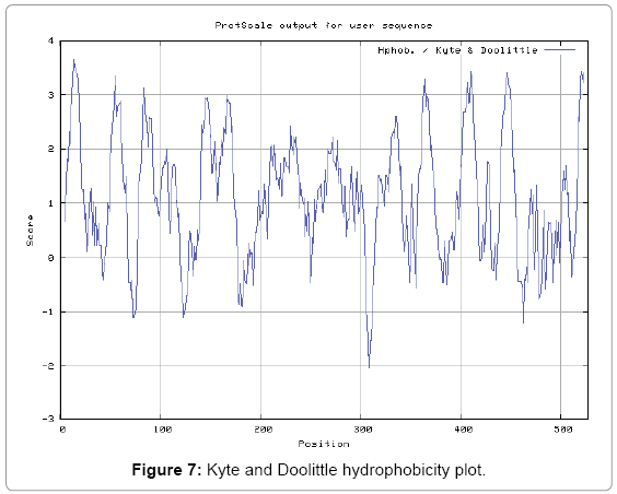
Figure 7: Kyte and Doolittle hydrophobicity plot.
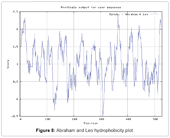
Figure 8: Abraham and Leo hydrophobicity plot.
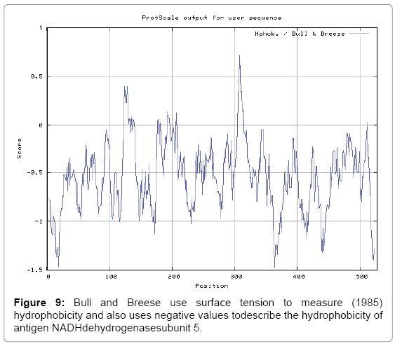
Figure 9: Bull and Breese use surface tension to measure (1985) hydrophobicity and also uses negative values todescribe the hydrophobicity of antigen NADHdehydrogenasesubunit 5.
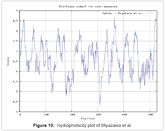
Figure 10: Hydrophobicity plot of Miyazawa et al.

Figure 11: Hydrophobicity plot of Roseman (1988).
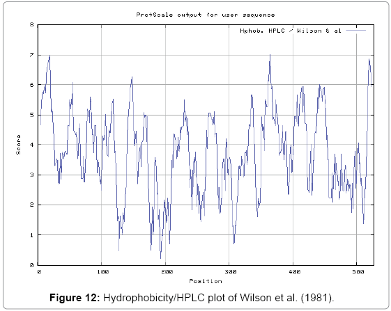
Figure 12: Hydrophobicity/HPLC plot of Wilson et al. (1981).
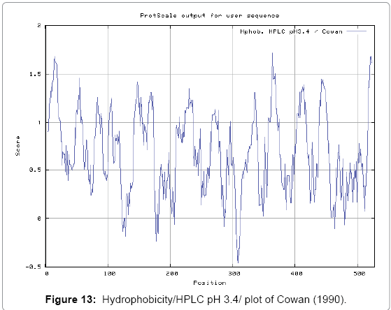
Figure 13: Hydrophobicity/HPLC pH 3.4/ plot of Cowan (1990).
An antigenic protein NADH dehydrogenase subunit 5 from D. medinensis might plays an important role in vaccine development. In future study the peptide fragments of antigen protein of NADH dehydrogenase subunit 5 can be used to select monomer for use in rational vaccine design and can develop the understanding of roles in the immune system in infectious disease.