Journal of Clinical and Experimental Ophthalmology
Open Access
ISSN: 2155-9570
ISSN: 2155-9570
Clinical Images - (2022)Volume 13, Issue 2
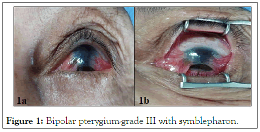
A 65-year-old patient presented with a pterygium with low visual acuity in the left eye. The clinical examination reveals that the patient's visual acuity was reducing to Counting Fingers (CF). The examination of the anterior segment showed a grade III bipolar pterygium with symblepharon.
An Optical Coherence Tomography (OCT) of the anterior segment showed a stromal infiltration of the pterygium. The surgical procedure consisted of a pterygium excision with the application of 5-Fluorouracil (5-FU) and a symplepharon release. A pterygium is a benign conjunctival fibro-vascular lesion. It’s encroaching progressively on the cornea and can become disabling [1]. The destruction of the limbus during the limbal stem cell deficiency involves a loss of its functions: The corneal epithelium can no longer regenerate by destruction of the limbal stem cell contingent and the limbic barrier can no longer contest against conjunctivalization [2,3] (Figure 1).

Figure 1:Bipolar pterygium-grade III with symblepharon.
[Crossref] [Google Scholar] [Pubmed]
[Crossref] [Google Scholar] [Pubmed]
Citation: Bezza H, Algouti Z, Mounsif A, El-Filali EM, Bouabbadi S, Zoaui K, et al. (2021) Study on Limbal Stem Cell Deficiency. J Clin Exp Ophthalmol. 13:910
Received: 08-Dec-2021, Manuscript No. JCEO-22-12922; Editor assigned: 10-Dec-2021, Pre QC No. JCEO-22-12922 (PQ); Reviewed: 24-Dec-2021, QC No. JCEO-22-12922; Revised: 29-Dec-2021, Manuscript No. JCEO-22-12922 (R); Published: 05-Jan-2022 , DOI: 10.35248/2155-9570.22.13.910
Copyright: © 2021 Bezza H, et al. This is an open-access article distributed under the terms of the Creative Commons Attribution License, which permits unrestricted use, distribution, and reproduction in any medium, provided the original author and source are credited.