Anatomy & Physiology: Current Research
Open Access
ISSN: 2161-0940
ISSN: 2161-0940
Review Article - (2024)Volume 14, Issue 3
Introduction: Digital anatomy uses new multimedia and Three-Dimensional (3D) tools. In Senegal, this teaching approach is less widespread. The aim of our study is to evaluate the content and impact of the use of Virtual Dissection Table (VDT) in the development phase in the learning of osteology.
Methodology: An analytical cross-sectional study is conducted with presenters and students enrolled in first year of medical study at Cheikh Anta Diop University in 2021-2022. The practical work sessions of osteology take place with the articulation of two methods: Traditional classical and digital. This required the use of VDT in addition to other conventional equipment.
Results: For 93.6% of students, the explanations were given using the VDT were clear or very clear. In 94.1% of cases, they were satisfied or very satisfied with the illustration sequences performed with the VDT. It helped 91.6% of students to understand and assimilate osteology. The 93.6% of the students preferred to work with the VDT rather than without it. The presenters awarded an average of 2/10 for the accessibility of cadaver dissection, with extremes of 0/10 and 5/10. Graphic quality received an average of 5.45/10 and anatomical accuracy 5.09/10. The 81.8% of presenters preferred to use the VDT during osteology practical work sessions.
Conclusion: The development of Digital Anatomy does not rely solely on hardware resources. It also requires the expertise of the Anatomist during the development process of the digital tool.
Digital anatomy; Virtual dissection table; 3D modeling; 3D printing
Human anatomy is the study of the structure and shape of the human body and the relationships between its various organs. It is an essential discipline for doctors at all stages of their training, from initial to continuing, and throughout their practice [1].
Digital anatomy is a new didactic approach to human anatomy, using new multimedia and three-dimensional (3D) tools.
In the West, these multimedia and 3D tools are flourishing, and are becoming increasingly important in teaching and research. Their integration and application in the teaching of lectures and practical work in anatomy have propelled digital anatomy as a new didactic approach. Lecture theaters and classrooms have been renovated and equipped with high-tech tools. Students also have access to the new tools themselves, but in pocket format. Also, 3D anatomical models of the female pelvis built by expert anatomists have provided answers about the complex structure and organization of the organs contained in this region [2].
In Africa, digital anatomy already used by some universities and has already been evaluated. According to authors Benleghib, et al., most students who participated in their studies found the use of digital anatomy excellent [3,4].
In Senegal, however, the use of digital technology to teach anatomy is less widespread. Digital anatomy has yet to take its rightful place in medical training establishments. This, despite the obstacles that have been erected to the classic or traditional didactic approach to Anatomy. Indeed, dissection, a technique that allows students with purely theoretical knowledge to be confronted in practice with the reality and complexity of the human body is not currently feasible during initial medical training [5,6]. It is important to recognize that in Senegalese society, the sacred mental image of the human body dating back to antiquity still prevails. The human body arouses fear and prohibitions in the minds of most of the general population. Cultural, social, and religious representations make didactic dissection almost impractical for students. Body donation is still non-existent. Legal provisions for using autopsy bodies for scientific purposes are not up to date, and families are fiercely on guard to verify the integrity of the bodies of their deceased loved ones.
On the other hand, our institutions are confronted with overcrowding. The average number of first-year medical students over the last two years is around 700. With this number of students, it will not be possible to provide them with enough anatomical autopsy specimens or cadavers.
This situation may well justify the imminent use of virtual dissection, which will free us from the ethical concerns associated with body dissection [6]. Virtual dissection uses a removable touch screen on which 3D images are displayed, allowing anatomical structures to be visualized through different planes and slice levels. Thus, at the Dakar Laboratory of Anatomy and Organogenesis, the acquisition of a Virtual Dissection Table (VDT) during the 2021-2022 academic year, enabled us to observe that its use could facilitate the acquisition (learning speed and quality) of skills for learners in Human Anatomy given the real obstacles facing cadaver dissection.
The aim of our study is therefore to evaluate the content and impact of using VDT in the development phase in the learning of Osteology.
Study setting
The teaching experiment was carried out in the Anatomy and Organogenesis Laboratory of the Faculty of Medicine, Cheikh Anta Diop University, Dakar (Figures 1 and 2).
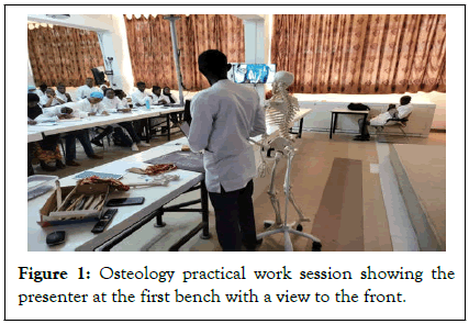
Figure 1: Osteology practical work session showing the presenter at the first bench with a view to the front.
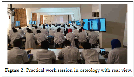
Figure 2: Practical work session in osteology with rear view.
Type of study and study period
Our study was a cross-sectional analytical study. The form was opened over the period January 2023 to March 2023.
Study subjects
The study focused on presenters and students enrolled in first year Bachelor of Medicine for the academic year 2021-2022.
Inclusion criteria
The following were included: 1) Students who consented to the study, 2) Students with a maximum of two enrolments in first year Bachelor of Medicine, 3) Presenters having consented to the study, 4) Presenters with at least 3 experiences of using the VDT.
Exclusion criteria
The following were excluded: 1) Students who did not answer the required questions on the form, 2) Presenters who had experienced VDT less than 3 times.
Materials
Materials that are utilized are:
Building and characteristics: The 3D mesh models of the male and female were built in the Paris Cité University from the anatomical slices of the korean visible human [7-9]. A dedicated software was built with Acrobat Pro interface in JavaScript to manipulate the 3D models using. U3d format. The hardware is a mac minicomputer with 16 gigas of Ram connected to a 55 inches’ touch monitor (Figure 3).
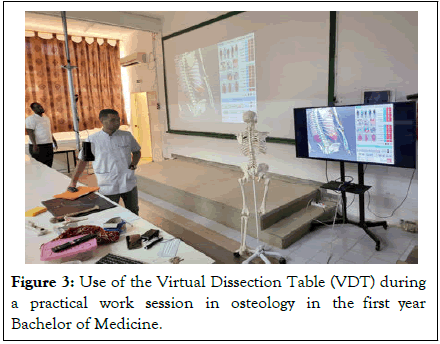
Figure 3: Use of the Virtual Dissection Table (VDT) during a practical work session in osteology in the first year Bachelor of Medicine.
Conventional and digital media: Nine projection screens, a video projector, a camera for the presenter, real and synthetic human bones, chalk, and a black file folder converted into a mini blackboard (Figure 4).
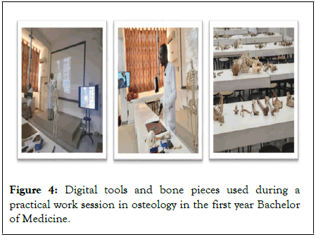
Figure 4: Digital tools and bone pieces used during a practical work session in osteology in the first year Bachelor of Medicine.
Methods
A few of the methods include:
Session preparation: One week prior to each practical work session, we proceeded as follows: 1) Meeting with the team of presenters, made up of Anatomy instructors, part-time teachers, and the full-time teacher in charge of practical work (Figure 5), 2) Selection of illustration sequences from the contents of the VDT, based on the quality of the 3D rendering of the anatomical structures shown and their relevance to the bone to be presented, 3) Insertion of selected and validated illustration sequences into the overall presentation, 4) Validation of the scenario constructed using real bone parts, the usual digital supports and VDT, 5) Rehearsed simulation of the session by presenters from the established osteology practical and practical training program.
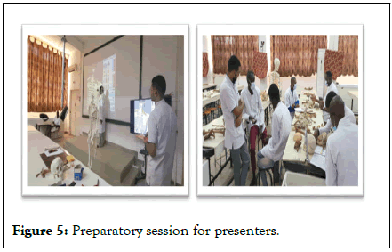
Figure 5: Preparatory session for presenters.
Session sequence: Practical work sessions last 1 hour. The session comprises 3 successive stages: 1) A 35-minute stage during which the presenter manipulates and describes the bones with direct camera transmission on 8 TV screens. Students, especially those occupying the benches at the back of the room, follow along with the bones in hand and the TV screen (Figure 6), 2) This is followed by a second 10-minute stage in which the presenter manipulates the bone studied in 3D and illustrates clinical applications using VDT (Figure 7), 3) A 3rd 15-minute stage is given over to the students, who reproduce a bone handling sequence on the benches and ask questions (Figure 8).
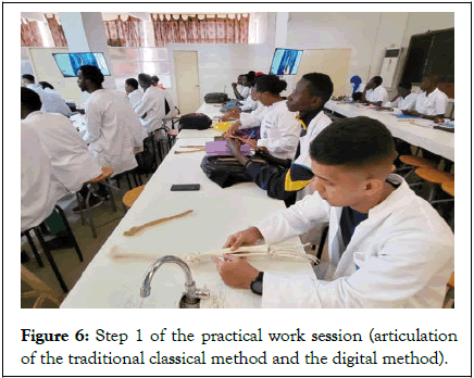
Figure 6: Step 1 of the practical work session (articulation of the traditional classical method and the digital method).
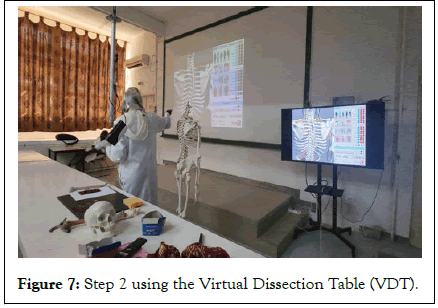
Figure 7: Step 2 using the Virtual Dissection Table (VDT).
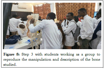
Figure 8: Step 3 with students working as a group to reproduce the manipulation and description of the bone studied.
Data collection
We proceeded with consecutive, non-exhaustive sampling. Posthoc feedback from students and presenters was collected in the form of an electronic questionnaire using Google form.
Students informed the following fields on the form: 1) Sociodemographic characteristics, 2) The clarity of the presenters' explanations when using the VDT, 3) Overall assessment of the VDT illustration sequences, 3) The contribution of VDT to the understanding and assimilation of osteology, 4) L1 students' preferences for using the VDT, 5) Students’ experience in using virtual dissection software.
The presenters informed the following fields: 1) Sociodemographic characteristics, 2) Presenters' experience and opinion of cadaver dissection, 3) Presenters' scorings of VDT in the development phase, 3) Presenters' general assessment of VDT, 4) Presenters' preferences for using the VDT, 5) Presenters' experience with virtual dissection software.
Operational definition of study variables
Students' assessment of the clarity of the presenter's explanations when using the VDT was assessed using a 4-item Likert clarity scale: Incomprehensible, unclear, clear, and very clear. The overall assessment of illustration sequences using the VDT was evaluated using a Likert satisfaction scale, consisting of 4 items: very unsatisfied, unsatisfied, satisfied, and very satisfied.
Statistical analysis
Data were analyzed using SPSS (Statistical Package for Social Sciences) version 25.0. Tables and graphs were produced using Microsoft Excel 2020. Quantitative variables followed a nonnormal distribution and were represented by the median and the interquartile range. In some cases, they were grouped into classes. Qualitative variables were represented either by a frequency table or a pie chart. The Spearmann test was used to determine whether there was a correlation between the score given by students to assess the clarity of explanations and that given to assess their overall assessment. The Chi-2 test and Fischer's exact test were used in bivariate analysis to investigate factors that could potentially influence student satisfaction with the VDT. Any confounding factors were controlled for by multivariate analysis using binary logistic regression. The threshold of statistical significance was set at 5%.
Ethical considerations
To conduct this study, we obtained the approval of the Anatomy Chair of the Faculty of Medicine in Cheikh Anta Diop University. All data was collected considering compliance with the rules of medical ethics. Students and presenters were informed about the procedures and objectives of the study. The study protocol was designed and carried out in accordance with the recommendations of the Declaration of Helsinki. The data were stored by the study principal investigator in the Anatomy laboratory computer with secure access.
Student assessment of the VDT in the development phase
A total of 366 students enrolled in first year Bachelor took part in the study.
Socio-demographic characteristics of the study population (n=366): The students who took part in this study were aged between 18 and 25, with an estimated median age of 20. 70% of them were between 20 and 21. There was a slight male predominance, with a sex ratio of 1:19.
Evaluation of first year Bachelor students' participation in practical work sessions (n=366): Among the students, 98.1% participated in the osteology practical sessions with the VDT. Validation of the exam in the previous year, late orientation to Medicine and late entry to the laboratory explain the overall non-participation of the other students.
Students' perception of the clarity of explanations: Students gave a median score of 8/10 for the clarity of explanations. Thus, for 93.6% of them, the explanations given when using the VDT were clear or very clear.
Overall assessment of the 3D illustration sequences used: Students also gave a median score of 8/10 for their overall assessment of illustration sequences with the VDT. In fact, 94.1% of them were satisfied or very satisfied with the illustration sequences created with the VDT.
Students’ perceptions on the contribution of the VDT to understanding and assimilation of osteology: The VDT helped 91.6% of first year Bachelor students to understand and assimilate well osteology.
Students' preferred use of the VDT for practical and homework sessions: The 93.6% of students preferred to use the VDT for practical work sessions, rather than without it.
Students' experience of using virtual dissection software: Prior to the practical sessions with the VDT, only 23.7% of first year Bachelor students were already using virtual dissection software.
Correlation between clarity of explanations and overall assessment of illustration sequences: We found a very strong positive correlation (r=0.81) between the clarity of explanations when using VDT and the students' overall assessment of the illustration sequences (Figure 9).
Figure 7: Step 2 using the Virtual Dissection Table (VDT).
Figure 9: Correlation between the clarity score of the explanations and the overall assessment score of the illustration sequences (Spearmann correlation test). NOTE: P<0.001, r=0,81.
Factors influencing overall assessment of illustration sequences using the VDT: In bivariate analysis, students' preference for using the VDT during practical works and the clarity of the explanations provided by the presenters seemed to be the main determinants of their satisfaction (Table 1).
| Factors | Overall appreciation | OR (CI 95%) | p-value | |
|---|---|---|---|---|
| Satisfied | Unsatisfied | |||
| Age | ||||
| <20 years | 78 | 3 | 1.80 (0.51-6.27) | 0.43 |
| ≥ 20 years | 260 | 18 | ||
| Sex | ||||
| Male | 186 | 13 | 0.75 (0.30-1.86) | 0. 65 |
| Female | 152 | 8 | ||
| Student's preference | ||||
| With 3D VDT | 17 | 6 | 0.13 (0.05-0.38) | 0.001 |
| Without 3D VDT | 321 | 15 | ||
| Previous use of 3D software | ||||
| Yes | 77 | 8 | 0.47 (0.19-1.19) | 0.11 |
| No | 261 | 13 | ||
| Clarity of explanations | ||||
| Clear | 328 | 8 | 53.3 (18.05-157.32) | <0.001 |
| Not clear | 10 | 13 | ||
Table 1: Factors influencing overall appreciation of illustration sequences performed with the VDT: Bivariate analysis using the Chi-2 Test and Fischer's Exact Test.
Factors influencing overall assessment of illustration sequences using the VDT: After multivariate analysis using binary logistic regression and controlling for confounding factors, we found that only the clarity of the explanations given by the presenter to the students when using the VDT determined their overall assessment of the illustration sequences performed during the practical work (p<0.001) (Table 2).
| Factors | Overall appreciation | OR aj (CI 95%) | p-value | |
|---|---|---|---|---|
| Satisfied | Unsatisfied | |||
| Preference to use 3D TDVD | ||||
| With table | 17 | 6 | 0.25 (0.06-1.08) | 0.06 |
| Without table | 321 | 15 | ||
| History of software use | ||||
| Yes | 77 | 8 | 0.74 (0.23-2.41) | 0.62 |
| No | 261 | 13 | ||
| Clarity of explanations | ||||
| Clear | 328 | 8 | 41.78 (13.52-129.13) | <0.001 |
| Unclear | 10 | 13 | ||
Table 2: Factors influencing overall appreciation of illustration sequences using the 3D VDT (Multivariate analysis using binary logistic regression).
Presenters' assessment of VDT in the development phase.
Number of participants: There were 11 presenters.
Socio-demographic characteristics of presenters: The average age was 36.45, with extremes of 29 and 45. Men represented 72.7% and women 27.3%.
Function of presenters: Both tenured and part-time teachers responded to the form, with a proportion of 36.4% each. Monitors accounted for 27.2%.
Cadaver dissection: The presenters gave an average of 2/10 for the accessibility of cadaver dissection, with extremes of 0/10 and 5/10 (Table 3).
| Rating categories | Mean/10 | Standard deviation | Minimum score/10 | Maximum score/10 |
|---|---|---|---|---|
| Score for accessibility to cadaver dissection | 2 | 1.55 | 0 | 5 |
Table 3: Presenters notes on the accessibility of cadaver dissection.
Most presenters (72.7%) had already experienced cadaver dissection. Their opinion of cadaver dissection was 54.5% favorable. However, 81.8% felt that virtual or digital dissection was a good alternative to cadaver dissection in the absence of a cadaver (Table 4).
| Variable | Number | Percentage (%) |
|---|---|---|
| Previous performance of dissections on cadaver | ||
| Yes | 8 | 72.7 |
| No | 3 | 27.3 |
| Opinion regarding cadaver dissection | ||
| Favorable | 6 | 54.5 |
| Very favorable | 1 | 9.1 |
| Mixed | 1 | 9.1 |
| Not answered | 3 | 27.3 |
| Virtual dissection as a good palliative in the absence of a body | ||
| Yes | 9 | 81.8 |
| No | 2 | 18.2 |
| Total | 11 | 100 |
Table 4: Experience and opinion of presenters on cadaver dissection.
Presenters' scorings of VDT in the development phase: The presenters awarded an average of 5.91/10 for the ease of use of the VDT and 5.64/10 for the simplicity of the interface. For content, graphic quality was given an average of 5.45/10 and anatomical accuracy 5.09/10. They awarded scores of 6.09/10 and 5.27/10 respectively for quizzes and overall assessment of the VDT (Table 5).
| Score categories | Mean/10 | Standard deviation | Minimum score/10 | Maximum score/10 |
|---|---|---|---|---|
| Score for ease of use | 5.91 | 1.87 | 2 | 8 |
| Score for interface simplicity | 5.64 | 1.75 | 3 | 9 |
| Score for graphics quality | 5.45 | 1.51 | 3 | 8 |
| Score for anatomical accuracy | 5.09 | 1.81 | 1 | 7 |
| Score for Quizzes | 6.09 | 1.97 | 2 | 9 |
| Score for overall assessment | 5.27 | 1.74 | 2 | 8 |
Table 5: Rating of the 3D TDV by the presenters.
Functionalities to be added: The presenters suggested functionalities on 3D model animation at 9.1% and on the use of dissection videos at 9.1%.
Preferences for practical work with or without VDT: The 81.8%of presenters preferred to use VDT for osteology practical work sessions.
Previous experience with virtual dissection software: Previous experience with virtual dissection software was 54.5% (Table 6).
| Variables | Number | Percentage (%) |
|---|---|---|
| Previous use of virtual dissection software | ||
| Yes | 6 | 54.5 |
| No | 5 | 45.5 |
| Total | 11 | 100 |
Table 6: Previous use of virtual dissection software by stakeholders.
Limitation to the study
The students answered the questions on the form after passing the practical Anatomy exam in their first year of medical school. They had therefore received their exam marks in Anatomy. Thus, the awarding of certain low scores for the evaluation of the DVT, such as 0/10, or answers such as "I don't know" to justify its absence, may be linked to frustration following a poor examination mark received by the student. However, this does not apply to presenters. We may suggest that, for other studies evaluating teaching tools, the data could be collected before the exam or before the exam marks are handed in. However, this could bias the evaluation marks that the students could award in the direction of complacency and bias the student's consent to participate freely in the study.
Preparing the Anatomy practical work session
The preparation of the practical work session is de facto required and requires supervision by a tenured teacher for the construction and validation of the scenario. It also has the advantage of standardizing the content to be transmitted to students, since years of experience vary from one presenter to another. Similarly, content that has been studied, validated, and standardized upstream by the teaching team reduces the likelihood of students getting lost when faced with the diversity and multitude of information sources on the Internet.
Furthermore, as the VDT used is still in the development phase, it is imperative during this preparatory session to assess the accuracy of the 3D rendering of its anatomical content. This is followed by the selection of validated structures, i.e., those with the best rendering close to reality.
Other structures with less accurate 3D rendering could be the subject of further research.
Advantages of combining two methods: Traditional classical and 3D digital
Presenters need to be able to combine the traditional classical method with the 3D digital one, to avoid technical problems that can arise at any time during digital anatomy sessions. These can include cable, touchscreen or projection malfunctions, or power cuts. It makes sense to convert the current stage of the presentation back to the traditional classical method. This involves rearranging the layout of the students around the benches and redeploying the other presenters to the sub-groups of students that have been formed. From then on, the session continues with this new organization, reflecting the presenters' mastery of both teaching methods.
Student satisfaction with VDT illustration sequences
The gold standard in practical anatomy work is cadaver dissection [1], and anatomists agree on this. Nevertheless, the feasibility and accessibility of this teaching method is difficult, as our results show. In Senegal, undergraduate medical students do not benefit from cadaver dissection practice. Cultural and religious barriers, the absence of legal provisions for the donation of corpses for scientific purposes, and overcrowding justify this inaccessibility to cadaver dissection.
Added to this is the problem of the laboratory's real bone heritage, which is currently undergoing significant deterioration. Several bone pieces have disappeared or been damaged, and there has been no subsequent restoration of this heritage.
The use of a VDT has made up for these real shortcomings, offering not only manipulable 3D images of bones, but also the ability to appreciate in 3D the muscular, membranous, and vascular-nervous relationships of the bones studied. This enables students to easily understand the clinical consequences of trauma to the bone. It also provides a close-up view of other soft tissues that cannot be visualized on the real bone parts usually used, or that are only appreciated in 2D. The most telling example is the study of nerve relationships in the humerus, the main bone of the upper arm. The passage of the radial nerve on the posterior surface of the shaft and that of the ulnar nerve on the posterior surface of the medial epicondyle are well illustrated by 3D visualization with VDT. The same is true of the interosseous membrane, which inserts at the interosseous edges of the two forearm bones. By using the 3D image from the VDT, the student is given a perfect mental representation of the situation and insertion zones of this interosseous membrane in the living subject. This was only possible with dissection, which has now become inaccessible in the Senegalese context.
This clear advantage of the VDT is confirmed by the results of our study, which showed that 94.1% of students were satisfied or very satisfied with the illustration sequences produced with the VDT. However, the preparatory practical work session could be decisive in the procedure for selecting appropriate anatomical content, accurate and with a 3D rendering as close to reality as possible. In this way, the positive result obtained regarding satisfaction with the VDT illustration sequences can also be partly attributed to the preparatory session for presenters.
Students' preferred use of VDT
In addition to their satisfaction, almost all the students expressed a preference for hands-on, directed teaching in which the VDT would be one of the pedagogical tools. However, in the study by Boukabache, et al., only 16.25% preferred a combination of the two methods, i.e., models and digital table [4]. The 83.75% of students found the digital dissection table, used on its own, to be the best medium for teaching anatomy [4].
In our study, 23.7% of first year Bachelor students were already using virtual dissection software. This result testifies to the need for anatomists to contribute to, and become involved in, the production of digital teaching tools to satisfy the demand formulated by these students. So, the path towards digital is no longer an option but rather a necessity.
Assessment of VDT content in the development phase
The presenters gave a fair average score for the content of the VDT in the development phase. In the study by benleghib. The quality of the animated images presented on slides was judged to be excellent [10]. However, this assessment was made by the students, whereas in our study it was the presenters who evaluated the content of the VDT.
These results point to the need for further work on the 3D reconstruction of the anatomical content of the VDT this will require greater precision and a better 3D rendering, as close as possible to reality.
Despite the average marks they gave, presenters preferred practical work sessions with VDT at 81.8%. Students also preferred practical work sessions with VDT at 93.6%. What's more, VDT helped 91.6% of students to fully understand and assimilate the illustration sequences. This testifies to the significant advantages of VDT, namely the 3D view of anatomical structures, which enables students to form a better mental representation of the structure under study. Added to this is the interactivity between presenters and students, which motivates the latter and keeps them attentive throughout the session. Delmas, et al., point out that digital anatomy facilitates interactivity between teachers and students [1].
This interactivity is enhanced by the availability of useful image manipulation functions. These include structure transparency and masking, as well as screen tactility and rotational movement. The ability to visualize structures internally, and to cut the body into different planes and levels, remains as relevant as ever, if not more so, when compared to conventional 2D images.
However, we can assume that the appropriation of VDT by both students and presenters was facilitated by the preparatory sessions of the scenario, the experience of the presenters and the articulation of the two traditional classical and digital methods.
The development of digital anatomy does not rely solely on material resources. It also requires the expertise of the Anatomist during the development process of the digital tool, the capitalization of skills from both traditional and digital teaching methods, and multidisciplinary collaboration with bioinformaticians and biomathematicians. ongoing research and development efforts should be supported to continually refine and expand the capabilities of digital anatomy tools. This includes integrating advancements in medical imaging technology, such as MRI and CT scans, to enhance the accuracy and detail of anatomical representations. Ultimately, the evolution of digital anatomy is a multifaceted endeavor that requires a holistic approach, combining technological innovation, educational expertise, and interdisciplinary collaboration to shape the future of anatomical education and medical practice.
[Crossref] [Google Scholar] [PubMed]
[Crossref] [Google Scholar] [PubMed]
Citation: Wade R, Thiam SAG, Garba KY, Ndiaye M, Tireira A, Seck ID, et al. (2024) Anatomical Expertise to the Development of Digital Teaching Tool. Anat Physiol. 14:481.
Received: 22-Apr-2024, Manuscript No. APCR-24-30888; Editor assigned: 24-Apr-2024, Pre QC No. APCR-24-30888; Reviewed: 08-May-2024, QC No. APCR-24-30888; Revised: 15-May-2024, Manuscript No. APCR-24-30888 (R); Published: 22-May-2024 , DOI: 10.35248/2161-0940.24.14.481
Copyright: © 2024 Wade R, et al. This is an open-access article distributed under the terms of the Creative Commons Attribution License, which permits unrestricted use, distribution, and reproduction in any medium, provided the original author and source are credited.