Journal of Clinical & Experimental Dermatology Research
Open Access
ISSN: 2155-9554
ISSN: 2155-9554
Case Report - (2020)Volume 11, Issue 4
Hobnail Hemangioma is a rare entity, first described by Santa cruz and Aronberg in 1988, it’s a lesion of vascular
origin, probably lymphatic. The most common clinical feature is a solitary violaceous papule surrounded by a pale,
thin area and a peripheral ecchymotic ring, simulating a target.
We report a rare case of hobnail hemangioma.
Hobnail Hemangioma; Dermatology; Skin
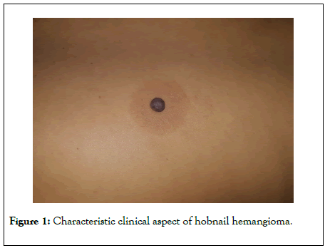
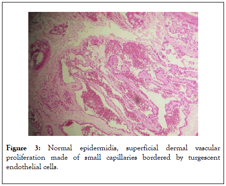
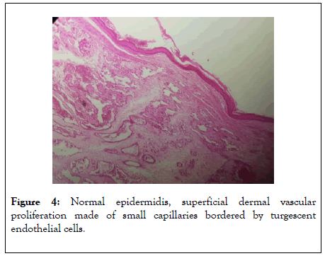
A 33-year-old woman, with no medical history, was visiting our dermatology’s department for an angiomatous lesion that did not disappear at the pressure, surrounded by a halo of pink skin and then a purpuric border that had been evolving for two years [1]. Surgical exeresis with histological analysis of the lesion foud under a normal epidermis, a superficial dermal vascular proliferation made of small capillaries with irregulars contours, bordered by turgescent endothelial cells without a mitotic figure. There was also an inflammatory lymphocytic infiltrate around the capillaries that dissected collagen fibers [2]. This vascular proliferation extended into the underlying derm where siderophags were visualized.
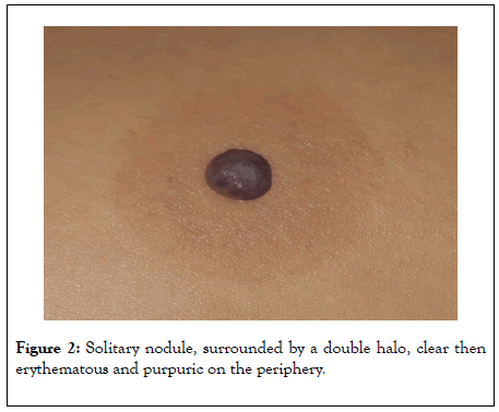
The very special clinical aspect of Hobnail Hemangioma makes it possible to evoke the diagnosis: it is a purple, solitary nodule, surrounded by a double halo, clear then erythematous and purpuric on the periphery, giving the lesion a target appearance [3,4]. Hobnail Hemangioma affects both the skin and mucous membranes without preferential topography. Some cases without targetoid formation have been reported [4].The peripheral purpuric halo can disappear spontaneously at the time of the thrusts. Hobnail Hemangioma is particularly common in adolescents and young adults.
The exact pathogenesis of THH is unknown, although it has been postulated that trauma is one of the main causes for the targetoid appearance [2,4,5,6] (Figures 1 and 2). Trauma could lead to the development of microshunts, in which the pressure of the capillaries would cause the filling of the lymph spaces of the lesion with erythrocytes, and contribute to the formation of aneurysmal microstructures [7]. The obstruction of some efferent lymphatic vessels would result in inflammation, fibrosis and interstitial hemosiderin deposits [5,7]. Hormones may also potentiate these tumors [8]. Histologically, it is a benign vascular proliferation made of small mature and superficial dilated vessels dissecting collagen bundles (Figure 3). Endothelial cells have a sparse cytoplasm and a prominent nucleus giving a characteristic appearance. The aspect of endothelial cells is reminiscent of epithelioid cells observed in epithelioid hemangiomes or angiolymphoid hyperplasias with eosinophilia. Peripheral erythematous or purpuric halo is a transient peripheral deposit of hemorrhage following hemorrhage on the periphery of the hemangioma [2,9,10]. Diagnosis is based on clinical-pathological correlation, especially of the typical morphology of the lesion. Simple excision is curative and allows a correct histological diagnosis [1,9] (Figure 4). Because it is a benign condition, such lesions are only removed for diagnostic or cosmetic reasons, since literature reports clearly indicate that neither local invasion nor dissemination take place. There are no reports of recurrence after excision of the lesion [9].

Figure 1: Characteristic clinical aspect of hobnail hemangioma.

Figure 2: Solitary nodule, surrounded by a double halo, clear then erythematous and purpuric on the periphery.

Figure 3: Normal epidermidis, superficial dermal vascular proliferation made of small capillaries bordered by turgescent endothelial cells.

Figure 4: Normal epidermidis, superficial dermal vascular proliferation made of small capillaries bordered by turgescent endothelial cells.
The diagnosis of HH is a rare entity, but not to be unknown by its very evocative clinical aspect.
No conflicts of interest
Citation: Belmourida S, Sialiti S, Meziane M, Ismaili N, Benzekri L, Hassam B, et al (2020) The Hobnail Hemangioma. J Clin Exp Dermatol Res. 11:528. DOI: 10.35248/2155-9554.20.11.528
Received: 11-Jul-2020 Accepted: 21-Jul-2020 Published: 28-Jul-2020 , DOI: 10.35248/2155-9554.20.11.528
Copyright: © 2020 Belmourida S, et al. This is an open-access article distributed under the terms of the Creative Commons Attribution License, which permits unrestricted use, distribution, and reproduction in any medium, provided the original author and source are credited.