Journal of Clinical & Experimental Dermatology Research
Open Access
ISSN: 2155-9554
ISSN: 2155-9554
Case Report - (2020)Volume 11, Issue 5
The retronychia (RN) is an incomplete form of nail shedding that leads to embedding of the nail into the proximal
nail fold and subsequent inflammation. It was first described by de Berker et al. 1999, "retro" meaning backwards and
"onychia" nail.
We report a new case.
Retronychia; Nails; Dermatology
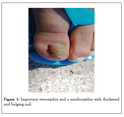
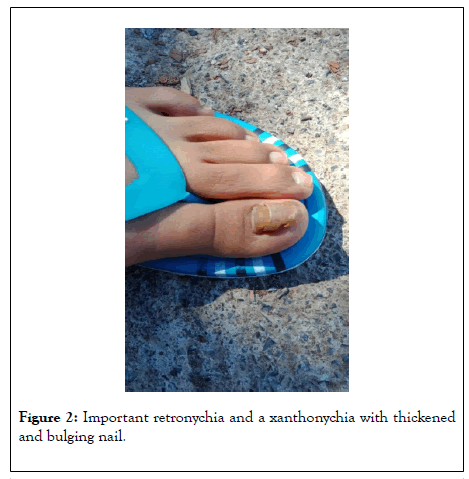
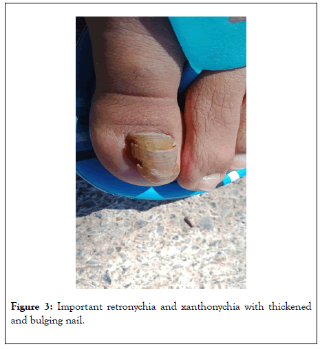
A 20-year-old girl, with no particular history, who presented to the dermatology department for chronic ungual hyperkeratosis of the left hallux evolving for 11 months associated with a discontinuation of nail growth [1]. There was no fever or initial trauma. On clinical examination, we had objectified, a xanthonychia with thickened and bulging nail, and there was no proximal perionyxis, sub-ungual inflammatory or exudation.
The diagnosis was a RN. The avulsion of the tablet confirmed the diagnosis with five generations of superimposed nails.
RN is rare, a few cases have been reported in the literature [2-9]. Trauma or systemic illness are the most common precipitating events, causing disruption of longitudinal nail bed growth and vertical growth of the new plate into the proximal nail fold. The condition is associated with multiple generations of nail plate misalignment beneath the proximal nail [10]. RN is a disease of adulthood predominating in females, although it has also been reported in children and in eldery [8,10-12]. Bilateral involvement and retronychia in multiple digits have been reported [8,10,12-16]. Clinical suspicion of retronychia is based on an unresolved chronic proximal paronychia in the setting of disrupted nail growth and multiple generations of nail plate [8,12,16]. The presence of granulation tissue between proximal and lateral nail folds is common. The diagnosis is usually clinical. In beginner or questionable cases, high-resolution ultrasound can assist in diagnosis [2,3]. Differential diagnosis should include diseases that can present as chronic paronychia (psoriatic arthritis, candidiasis or bacterial infections), subungual tumors (Bowen's disease, keratoacanthomas, and amelanotic melanoma), and cysts [4,5,16,17]. For treatment, some minor forms may evolve spontaneously favorably, they must be treated first-line by the application of corticosteroids on the dorsal fold under occlusion [7]. For major forms, surgical avulsion is necessary. It confirms the diagnosis by highlighting adhesive and superimposed nails [6] (Figures 1-3).

Figure 1: Important retronychia and a xanthonychia with thickened and bulging nail.

Figure 2: Important retronychia and a xanthonychia with thickened and bulging nail.

Figure 3: Important retronychia and xanthonychia with thickened and bulging nail.
The characteristic clinical aspect allows knowledge of this rare entity with rapid management avoiding inadequate local care and unnecessary antibiotic therapy especially in case of associated perionyxis.
No conflicts of interest
Citation: Belmourida S, Palamino H, Khallayoune M, Meziane M, Ismaili N, Benzekri L, et al (2020) The Retronychia. J Clin Exp Dermatol Res. 11:529. DOI: 10.35248/2155-9554.20.11.529
Received: 11-Jul-2020 Accepted: 21-Jul-2020 Published: 28-Jul-2020 , DOI: 10.35248/2155-9554.20.11.529
Copyright: © 2020 Belmourida S, et al. This is an open-access article distributed under the terms of the Creative Commons Attribution License, which permits unrestricted use, distribution, and reproduction in any medium, provided the original author and source are credited.