Clinical Pediatrics: Open Access
Open Access
ISSN: 2572-0775
ISSN: 2572-0775
Case Report - (2021)Volume 6, Issue 11
ACC is a rare malformation characterized by solitary lesion involving various layers of the skin or subcutaneous tissue and sometimes the bone on the scalp at birth. The lesion less involving the limbs or abdomen, but mostly on the scalp. Herein, we report a case of absence of skin involving the scalp at birth, diagnosed on ACC.
Aplasia cutis congenita; Type Ⅰ; Newborn; B-scan ultrasonography
The child, a female infant from China with birth weight of 3,300 g and gestational age of 35+5 week to a 28-year-old Para 2 mother. Apgar score was 10/10 at 1 minute, amniotic fluid, umbilical cord and placentas were normal. The baby was admitted to Neonatal intensive care unit account of absence of part of skin on the head noticed at birth. Parents in non-consanguineous setting. It is a sign of heterogeneous group of situations characterized by the congenital absence of dermis, epidermis and in some circumstances, hypodermal tissues or bone generally involving the scalp vertex.
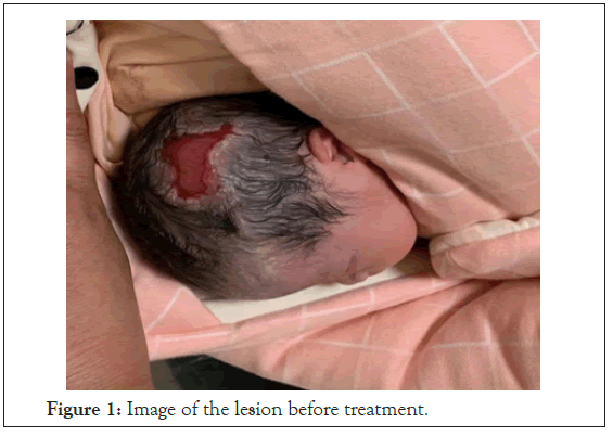
There was no drug use during pregnancy and no history of congenital anomalies in the extended family. There was no history of exposure to radiation in pregnancy. On physical examination, an irregularly shaped defect on the scalp over the temporal bone measuring 4 cm × 3 cm. A translucent layer of membrane covered the defect, vascular can be seen under the membrane and no obvious liquid exudation (Figure 1). There was no skin defect on the trunk and extremity, and no abnormalities of other organs. On laboratory examination, the results of a routine blood tests, liver and renal function, Echocardiography, abdominal B-ultrasound was normal. B-scan ultrasonography showed that the skull structure was complete. She has been diagnosed with ACC finally. This lesion was conservatively treated, including saline cleansing and application of Recombinant Bovine Basic Fibroblast Growth Factor gel twice a day. 25 day later, scar tissue has formed over the defect (Figure 2), the patient discharged. We followed the patient for two months. One month after discharged, the lesion where most of the scabs fell off has been healed (Figure 3). Two month after discharged, the lesion was healed completely, and there was no hair on the surface (Figure 4).

Figure 1: Image of the lesion before treatment.
Figure 2: Image of the lesion at discharged.
Figure 3: Image of the lesion one month after discharged.
Figure 4: Image of the lesion two month after discharged.
We ACC are a rare condition resulting in the absence of skin that can exist alone or combined with some types of genetic diseases [1]. Eldad reported that ACC occurring 3/10,000 live birth, this disorder is mainly in female [2]. The etiology of ACC is not clearly understood. Both mother’s exposure to toxins during pregnancy, intrauterine infection and placental vascular embolism have been implicated. ACC presenting with an aseptic tissue defect looks like an ulcer, and a thin, transparent and fragile membrane covered the defect that can involving various layers of the skin and sometimes the bone. Up to 86 percent of lesions are located on the scalp in the midline vertex position. Except for the ACC located on the scalp, this patient was not complicated with other abnormalities and belonged to Type I ACC (Scalp ACC without multiple anomalies) [3]. Its mode of inheritance is autosomal dominant or sporadic. This child has no family history and is considered to be sporadic.
The management of can be conservative surgical or a combination of both depending on the presentation, size and location of defect. The goal of conservative treatment includes reducing the risk for infection, providing protection, and promoting healing [4,5]. Most small areas of ACC can heal under conservative treatment [6-8]. In contrast, surgical treatment are necessary when the defected area is large and involves critical organs or tissues such as the scalp while also accompanied by the exposure of large blood vessels or sagittal sinus to prevent infection, and reduce the occurrence of complications. The conservative treatment is easy to carry out, simple, and allows granulation and complete curative so many trials, studies and clinical reports are recommended the Dura mater is accomplished of development of new bone. There some disadvantages in the conservative treatment those are Hemorrhage, Infections, Sagittal sinus thrombosis, and Hydrocephalus. While coming to the surgical treatment options include rotation flaps, skin grafts, free flaps, and tissue developments. When compare with the conservative treatment surgical is having some advantages like it reduces the risk of meningitis, sinus thrombosis, or hemorrhage. The disadvantages of the surgical treatment are Delayed healing, and Scares. In this case we take only the Type 1 ACC condition so mainly focus on the conservative treatment the subject we selected was cured in the procedure of the treatment because there are any other compilations are not involved with the new born. The treatment is succeeding in the case of this female new born. But there was no hair in the recovery portion of the scalp.
In this report, we presented a case with type I ACC, localized in the scalp of a female newborn. B-scan ultrasonography showed that the skull and meninges were not involved, and the result was good after conservative treatment. In this case surgical treatment is not required because of the size and condition of the Congenita. The treatment will depend upon the Newborn condition and Congenita condition.
Citation: Lin X, Min S, Wang D, Jiang MG (2021) Type I Aplasia Cutis Congenita: Case Report. Clin Pediatr. 6: 196
Received: 12-Nov-2021 Accepted: 26-Nov-2021 Published: 03-Dec-2021
Copyright: © Lin X, et al. This is an open-access article distributed under the terms of the creative commons attribution license, which permits unrestricted use, distribution, and reproduction in any medium, provided the original author and source are credited.