Internal Medicine
Open Access
ISSN: 2165-8048
ISSN: 2165-8048
Case Report - (2019)Volume 9, Issue 2
Werner syndrome is considered inherited premature ageing and genomic instability syndrome. It is an autosomal recessive disorder in which aging process is accelerated starting after puberty. It is also termed as Progeria adultorum.
The hallmark features are short stature, senile facies, scleroderma like skin (dry atrophic skin, mottled darkness, telangiectasia, sclerodactyly, and gangrene), cataract, hypogonadism, contractures of skin over joint, premature atherosclerosis, loss of subcutaneous fat and ulcers over feet and leg. Treatment of painful ulcers is difficult with increased risk of malignancy (fibro sarcoma in 10% of patients). Death is usually occurring in the fourth to sixth decade due to myocardial infarction or malignancy.
We reported 2 brothers of Pangeria of positive consanguinity parents the older is 40 years old, short stature, senile facies with bilateral hip joint replacement, chronic leg ulcer, bilateral cataract extraction and the younger is 35 years old, short stature, senile facies, infertile, previous cataract extraction with bone deformity. The later presented to us in outpatient clinic, Internal Medicine department, Tanta university Hospital, Egypt for preoperative assessment for cataract operation. We admit the patient to investigate him and to confirm our provisional diagnosis as regard Werner syndrome. As no definite treatment for this disorder and death usually occurs in the fourth to sixth decade, early diagnosis and follow up is beneficial and screening for malignancies and associated diseases should be performed regularly.
Werner syndrome; Pangeria; Scleroderma; Skin; Premature ageing; Bone deformity
Werner syndrome is an autosomal recessive disease that characterized by accelerated senility after puberty. It has a global incidence rate of less than 1 in 100,000 live births, but incidence in Japan is higher [1-4]. The mutation is in the WRN gene (Werner Syndrome RecQ like Helicase) that encode for DNA helicase which is thought to be important for the maintenance and repair of DNA. This mutation exhibits accelerated DNA methylation changes that are similar to those occurred in normal senility [5]. It causes genetic instability and failure of repair DNA.
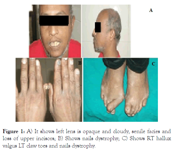
A 35-year-old married male presented to our unit for preoperative assessment for cataract operation. We noted that he have senile facies with partial loss of hair which started at the age of 20, short stature, dark skin, lost one of upper incisors, high pitched voice, bone deformity in toes of both foot for left big toe and he was operated at age of 30, with tight skin over feet, distal digital joint contractures and skin thickening which gradually worsened. History of bilateral cataract for one of them (Right eye) he was operated at the age of 31. Patient married since 10 years and has no children, no diabetes but hypertension was first discovered during admission (Figure 1).

Figure 1: A) It shows left lens is opaque and cloudy, senile facies and loss of upper incisors; B) Shows nails dystrophy; C) Shows RT hallux valgus LT claw toes and nails dystrophy.
He was born from a consanguineous marriage and his father and mother were normal. However, one of his siblings has similar disease.
On physical examination patient appeared much older than his stated age and weighed 49 kg. The height was 150 cm. The trunk was normal with distended abdomen, but the extremities were slender. The scalp hair was partially lost, greyish white, sparse, facial hair white. There was microstomia and the nose was small and beaked. The skin of the face was atrophied, wrinkled with little subcutaneous tissue but was not sclerosed. Contractures of hands and feet with mottled hyperpigmentation were present with 20 nail dystrophy. On examination of eyes, unilateral central lens opacity in left eye was seen. One of upper incisor tooth was missing, blood pressure was high (170/100) mm Hg.
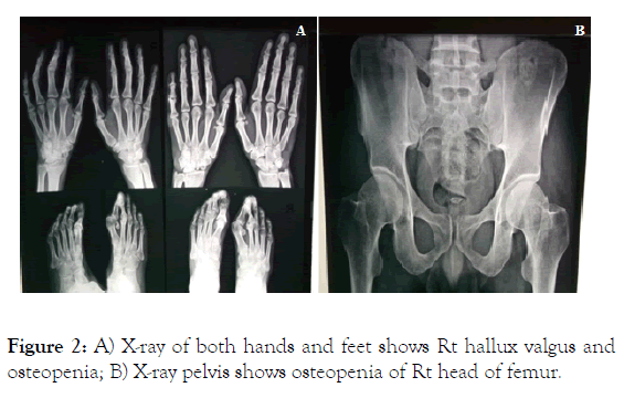
There were also crusted verucous plaques over heels and metatarsal bones. We ask the patient about similar condition in his family he clarified that his brother was operated for cataract extraction and hip joint replacement (Figure 2).

Figure 2: A) X-ray of both hands and feet shows Rt hallux valgus and osteopenia; B) X-ray pelvis shows osteopenia of Rt head of femur.
After admission of the patient, investigations showed in Table 1.
Table 1: Laboratory and radiological investigation.
| CBC | Mild normocytic normochromic anemia Hb: 12.8 g/dL, MCV: 84.7 fl, MCHC: 5.5%, PLT: 164000/cmm, WBCs: 8.500/cmm |
| Liver function test | Normal Total bilirubin: 0.5 mg/dL, SGPT: 28 µ/ml, SGOT: 30 µ/ml, S. albumin: 4.6 g/dL, Prothrombin activity: 83% |
| Kidney function tests | Normal Serum creatinine: 0.7 mg/dL, blood urea: 23 mg/dL |
| Serum electrolytes | Normal K: 4.2 mEq/L , Na: 140 mEq/L |
| Fasting blood glucose PPG | 98 mg/dL, 120 mg/dL |
| Lipid profile | Deranged Cholesterol: 231 mg/dL (reference value<200 mg/dL), serum triglyceride: 390 mg/dL (reference value <150 mg/dL) |
| FSH LH free Testosterone |
8.2 mIU/mL (N) 18.6 mIU/mL (H) 7.3 pg/mL (L) |
| TSH | 2.46 uIU/mL (N) |
| PTH, Ca++, phosphorus |
66.3 pg/mL (N) Serum ionized Ca (L): 3.7 mg/dL (reference value 4.4-5.2 mg/dL) S.phosphorus (N): 3 mg/dL |
| ESR | First hour 38 mm, second hour 70 mm |
| HCVAB | Negative |
| ECG | Sinus tachycardia |
| Echo-Doppler | Sclerotic aortic with mild aortic regurge with EF 72% |
| Chest X-ray | Free |
| abdominal US | Bilateral renal gravels |
| Neck US | Left small follicular cyst of thyroid measured 4×2.5 mm |
| Duplex on arterial system of lower limb | Showed atherosclerotic changes of arterial system |
| Testicular US | Shows bilateral atrophic testis with bilateral rim of hydrocele |
| Semen analysis | Shows no sperm |
| X-ray foot | Hallux valgus, Osteopenia |
DEXA scan
BMD of lumbar vertebrae shows normal measurements with average T-score of -0.7 (-9%), BMD (bone mineral density) of left femoral neck shows osteopenia with average T-score of -1.9 (-26%), BMD of the left radius and ulna shows osteopenia with average T-score of -1.7 (-14%).
On laryngeal examination there were Mobile vocal cords with patent airway and laryngitis (due to reflux).
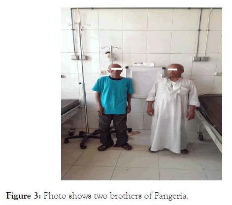
His brother is 40 years old, married and have one child (aged 5years), 145 cm, 40 kg, senile facies, dark skin, alopecia, small mandible, prominent zygomatic bone prominent eyes, high pitched voice, waddling movement due to shorting of one limb after recurrent hip joint replacement, the extremities were slender, tight skin over hands and feet, there was an dark, rough, punched out ulcer about 3 cm over the right lateral malleolus from 2 years ago not responding to usual medical treatment, history of bilateral cataract extraction with lenses insertion. His blood pressure was normal. Serum TG and cholesterol were moderately elevated (Figure 3).

Figure 3: Photo shows two brothers of Pangeria.
Unfortunately there is no definite cure for this syndrome but only prophylactic measures to avoid complication (cardiovascular and cerebral one) via treatment of hyperlipidaemia, osteoporosis and hypocalcaemia by alendronate and calcium supplement. Patient discharged from our unit after one week of admission and his blood pressure was controlled on ACEI (An Angiotensin-Converting-Enzyme Inhibitor) 25 mg/d, he was underwent cataract extraction with lens insertion and his brother referred for orthopedic consultation.
Pangeria is a very rare disorder; global incidence is 1 case in 1 million persons. It is recessive genetic disorder that is characterized by premature aging starting mostly after puberty and usually diagnosed in third decade [6]. It caused by mutation in the WRN gene that located on chromosome 8, position 12-11.2 [7]. The WRN gene is thought to be important for the maintenance and repair of DNA include replication, repair, recombination, transcription, and chromosomal isolation. Mutation causes genetic instability and failure of repair DNA. Cell cycle is primarily affected; patient undergoes very few cell divisions. It was known that the cells from normal individuals multiply approximately 60 times whereas Werner syndrome fibroblasts may reproduce only up to 20 times. Some papers have suggested that WRN is essentially a “counting gene,” regulating the total number of times that human cells may divide and reproduce. Brain and muscle cells not affected as they already undergo few cell division. The more individual ages, the less the cells undergo mitosis. They show signs of premature senility as well as having health complications of aging such as cardiovascular disease, cataract, premature menopause and bone deformity.
Considering the presented two brothers with Werner syndrome, we relatively diagnosed one patient at earlier age with respect to the published cases. Our diagnosis was based on the clinical findings and family history. We could not perform genetic study due to our laboratory condition. We would like to contribute to the previous literature with our clinical observation as regard the 2 brothers that the elder case is more manifested and complication is more evident (with difference of age 5 years from his brother) Generally, he was presented with alopecia, high pitched voice, extremity pain, recurrent hip joint replacement with abnormal movement due to shortage of one limb, scleroderma like skin in both foot and hand, chronic leg ulcer. They are still under follow up. Treatment is based on prophylactic measures to avoid complications.
Citation: Metwally S, El Ahwal L, Zaghlol K, Alwan N, Gabar R (2019) Werner Syndrome: A Case Report of Two Brothers of Pangeria. Intern Med. 9: 308. doi: 10.35248/2165-8048.19.9.308.
Received: 21-Jan-2019 Accepted: 20-May-2019 Published: 28-May-2019 , DOI: 10.35248/2165-8048.19.9.308
Copyright: © 2019 Metwally S, et al. This is an open-access article distributed under the terms of the Creative Commons Attribution License, which permits unrestricted use, distribution, and reproduction in any medium, provided the original author and source are credited.